Ovarian Cancer Ultrasound Images - Mage Of The Ovarian Tumor On Transvaginal Ultrasound Examination Download Scientific Diagram : If you have frequent or persistent symptoms of ovarian cancer, you will usually have a physical exam, including a pelvic exam.
Ovarian Cancer Ultrasound Images - Mage Of The Ovarian Tumor On Transvaginal Ultrasound Examination Download Scientific Diagram : If you have frequent or persistent symptoms of ovarian cancer, you will usually have a physical exam, including a pelvic exam.
Ovarian Cancer Ultrasound Images - Mage Of The Ovarian Tumor On Transvaginal Ultrasound Examination Download Scientific Diagram : If you have frequent or persistent symptoms of ovarian cancer, you will usually have a physical exam, including a pelvic exam.. Sagittal image of right ovary (sag rt, calipers). Clinical use of cancer biomarkers in epithelial ovarian cancer: Ovarian cancer is a relatively rare form of cancer that begins in the ovaries. An ovarian cancer ultrasound is one of several diagnostic tests that your doctor will use to determine if ovarian cancer is present. But these scans can't determine whether the abnormality is cancer.
1 nearly 75% of women with ovarian cancer present with advanced stage disease, which is associated with a poor prognosis. These small organs sit in a woman's pelvis and house the eggs that, when if ovarian cancer is suspected, you'll likely undergo a physical exam and imaging tests, such as an ultrasound or ct scan to look for tumors in the pelvic. Sound waves are released from a small probe placed in for ovarian cancer, the biopsy is most commonly done by removing the tumor during surgery. My mother found she had ovarian cancer when she was 50 years old. 2 there has been a relative standstill in the.
Ultrasound Imaging Of Ovarian Cancer Chapter 22 Ultrasonography In Gynecology from static.cambridge.org Ovarian cancer risk factors & prevention. An imaging test of the ovaries, such as a transvaginal ultrasound exam, may be done. If you're concerned that you may have symptoms of ovarian cancer, ask your gp about having a ca125 blood test and ultrasound scan. A type of ultrasound in which the device is placed in your vagina. The other ovary is not shown but showed a similar appearance. Ultrasound scans use high frequency sound waves to create a picture of a part of the body. Ultrasound scanners consist of a stand with a computer and electronics, a display screen to show the image. Sound waves are released from a small probe placed in for ovarian cancer, the biopsy is most commonly done by removing the tumor during surgery.
It started right after we were over.
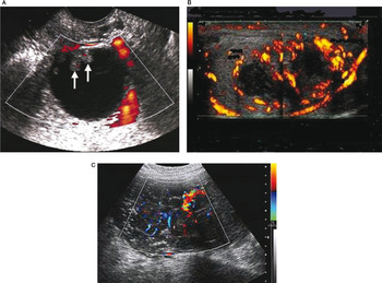
Genetic testing for ovarian cancer. Clinical use of cancer biomarkers in epithelial ovarian cancer: Imaging strategy for early ovarian cancer: This is yet another case of serous cystadenoma of the ovary (again it is difficult to determine the side of origin of this huge ovarian cyst from the ultrasound images above. The us images are of a young pregnant woman, who had multiple ovarian cysts. An imaging test of the ovaries, such as a transvaginal ultrasound exam, may be done. Sagittal image of right ovary (sag rt, calipers). Strength of the signalwhat instruments are used? Sound waves are released from a small probe placed in for ovarian cancer, the biopsy is most commonly done by removing the tumor during surgery. This ultrasound makes it possible for your doctor to obtain real time images of the current status of the ovaries as well as the rest of the reproductive system. The other ovary is not shown but showed a similar appearance. Ultrasound scans use high frequency sound waves to create a picture of a part of the body. For a vaginal ultrasound, they insert the probe into your vagina.
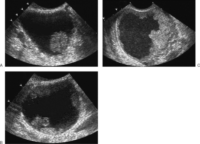
Ovarian torsion with no visible blood flow. A procedure used to examine the. If you have frequent or persistent symptoms of ovarian cancer, you will usually have a physical exam, including a pelvic exam. An ovarian cancer ultrasound is one of several diagnostic tests that your doctor will use to determine if ovarian cancer is present. During an external ultrasound of your pelvis, the doctor or radiographer moves a probe over the lower part of your tummy.
Ultrasound Evaluation Of The Adnexa Ovary And Fallopian Tubes Radiology Key from radiologykey.com Ovarian cancer is one of those nightmare cancers: Ovarian cancer is cancer that affects one or both ovaries. Characterization of adnexal masses with conventional and advanced imaging techniques. Department of health and human services national institutes of health. Ovary ultrasound education showing how to, scanning protocol, normal anatomy, anatomic variants, follicles and graafian follicle, corpus luteum. Ultrasound1 (ultrasonography) uses sound waves to create an image on a video screen. If ultrasound images show the former, further testing will be needed to diagnose cancer. If you're concerned that you may have symptoms of ovarian cancer, ask your gp about having a ca125 blood test and ultrasound scan.
Ovarian cancer images on ultrasound.
Imaging strategy for early ovarian cancer: Ovarian torsion with no visible blood flow. A radiologist—a physician who specializes in diagnosing. 1 nearly 75% of women with ovarian cancer present with advanced stage disease, which is associated with a poor prognosis. It started right after we were over. Br j obstet gynaecol 1990; To assess the value of ultrasonography in a screening procedure for early ovarian cancer. This is yet another case of serous cystadenoma of the ovary (again it is difficult to determine the side of origin of this huge ovarian cyst from the ultrasound images above. A procedure used to examine the. Department of health and human services national institutes of health. Current research suggests this cancer begins in the fallopian tubes and moves to the ovaries, the twin organs imaging tests, such as ultrasound or ct scans (seen here), can help reveal an ovarian mass. But these scans can't determine whether the abnormality is cancer. For a vaginal ultrasound, they insert the probe into your vagina.
A radiologist—a physician who specializes in diagnosing. A procedure used to examine the. Ovarian cancer is a relatively rare form of cancer that begins in the ovaries. This ultrasound makes it possible for your doctor to obtain real time images of the current status of the ovaries as well as the rest of the reproductive system. Ultrasound scans use high frequency sound waves to create a picture of a part of the body.
Ultrasound Evaluation Of The Adnexa Ovary And Fallopian Tubes Radiology Key from radiologykey.com The prognosis of ovarian cancer is poor compared with other gynaecological cancers. Ultrasound1 (ultrasonography) uses sound waves to create an image on a video screen. Ovarian cancer is the second most common gynecologic malignancy and is the fifth leading cause of cancer death in women. Ovarian cancer is a relatively rare form of cancer that begins in the ovaries. See more ideas about ovarian, ultrasound, ovaries. Strength of the signalwhat instruments are used? Ovary ultrasound education showing how to, scanning protocol, normal anatomy, anatomic variants, follicles and graafian follicle, corpus luteum. Ovarian cancer risk factors & prevention.
Ovarian cancer is one of those nightmare cancers:
Department of health and human services national institutes of health. The other ovary is not shown but showed a similar appearance. Imaging in gynecological disease 14 clinical and ultrasound characteristics of ovarian clear cell carcinoma pozzati 2018 ultrasound in obstetrics amp gynecology wiley online library. An imaging test of the ovaries, such as a transvaginal ultrasound exam, may be done. If you're concerned that you may have symptoms of ovarian cancer, ask your gp about having a ca125 blood test and ultrasound scan. Sagittal image of right ovary (sag rt, calipers). For a vaginal ultrasound, they insert the probe into your vagina. Ovary ultrasound education showing how to, scanning protocol, normal anatomy, anatomic variants, follicles and graafian follicle, corpus luteum. Of the various imaging methods that have been evaluated for use in ovarian cancer screening, transvaginal ultrasound has consistently proven to be the most effective technique. Characterization of adnexal masses with conventional and advanced imaging techniques. 1 nearly 75% of women with ovarian cancer present with advanced stage disease, which is associated with a poor prognosis. This ultrasound makes it possible for your doctor to obtain real time images of the current status of the ovaries as well as the rest of the reproductive system. Imaging strategies for ultrasound and mr imaging.
Characterization of adnexal masses with conventional and advanced imaging techniques ovarian cancer ultrasound. Of the various imaging methods that have been evaluated for use in ovarian cancer screening, transvaginal ultrasound has consistently proven to be the most effective technique.